
- About ACNS
- Meetings
- Annual Meeting and Courses
- Principles of Clinical Neurophysiology Courses
- Claim CME: Past Meetings
- Meetings & Programs Calendar
- Endorsed Courses & Symposia
- Education
- Practice
- Research
- Advocacy
- Membership
Contributed by
Dua’a Ba-Armah, MD and Cecil D. Hahn, MD, MPH, FACNS
The Hospital for Sick Children
Toronto, Canada
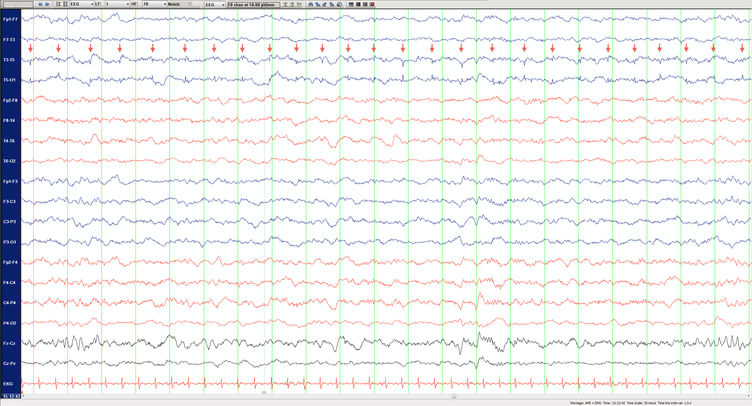
A 5-year-old boy with Down Syndrome presented with a history of intermittent episodes of eye rolling upwards or staring for few seconds with no postictal state. A routine outpatient EEG recording was obtained to assist with the diagnosis. The EEG, performed under sedation with chloral hydrate, revealed normal sleep features with superimposed intermittent periodic discharges in the left temporal head region (Figure 1), which occurred in sustained runs lasting between 30 seconds and 2 minutes. The discharges remained localized to the T5 electrode, maintained a consistent frequency of just over 1 Hz, and did not clearly evolve in frequency, amplitude or morphology. There was no associated focal slowing.
(click to enlarge)
Figure 1. Routine EEG in an A-P longitudinal bipolar montage displayed at 10μV/mm. FCz was used as a reference. The sampling rate was 200 Hz with 1-70 Hz band pass filter. Arrows illustrate periodic discharges phase reversing at T5.
What do these periodic discharges represent?
Because of the spiky morphology and the regular, periodic nature of the discharges without evolution in frequency, amplitude or morphology, an ECG or pulse artifact was considered, however the discharge frequency did not match the patient’s 120 bpm heart rate. Examination of the time-synchronized video recording revealed that the discharges appeared only when the child placed his head on his mother’s chest and disappeared promptly when the head was repositioned (Figures 2 & 3). Therefore, we concluded that these periodic discharges most likely represented an artifact generated by the mother’s heartbeat.
Artifacts are defined as unwanted electrical potentials originating from extra-cerebral sources.(1) Artifacts can be physiological or non-physiological.(2) Physiological artifacts include electrocardiographic artifacts such as pulse or ECG artifacts. Pulse artifacts are usually generated by minute movements of an EEG electrode due to pulsatile blood flow in an underlying scalp artery. Pulse artifacts are time-locked with the ECG, but they are usually slightly delayed with respect to the QRS complex due to the time it takes the systolic pulse of blood flow to reach the scalp. ECG artifacts are generated directly by the electrical field of the heart, which is oriented such that it produces a negative polarity signal on one side of the head and a positive polarity on the opposite side. For this reason, ECG artifact is particularly prevalent in referential montages with ear references (A1/A2).(2)
We considered the possibility that these periodic discharges represented either pulse or ECG artifact. The child’s head was wrapped with gauze, an electrical insulator, therefore it would seem more likely that pulsatile chest movement could have been transmitted to the T5 electrode rather than the direct electrical field of the mother’s electrocardiogram. However, the observed discharges had a spiky morphology more typical of ECG artifact than pulse artifact. It is possible that the head bandage was sufficiently loose to expose the T5 electrode, which could have come into direct contact with the mother’s chest covered only by a thin T-shirt, allowing for direct transmission of electrical potentials from the mother’s heart.
Identification and elimination of artifacts during the EEG recording are the responsibility of the EEG technologist. However, sometimes even the most expert technologists cannot eliminate all artifacts.(2) Artifacts remain one of the most common EEG findings. Correct identification of artifacts is vitally important so that they are not misinterpreted as a pathological finding. Occasionally, ECG artifact can partially obscure multiple channels of an EEG recording, making accurate interpretation difficult. Fortunately, algorithms are under development to permit filtering of such ECG artifacts.(3)

(click to enlarge)
Figure 2. Prompt disappearance of T5 rhythmic discharges when the child’s head is repositioned off the mother’s chest (blue arrow).

(click to enlarge)
Figure 3. Still image from time-locked video depicting position of the child’s head during the time of the periodic left temporal discharges.
References: